|
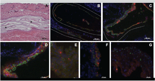
| Figure 5: Immunofluorescence analysis of characteristic urothelial markers. HE-stained cryostat section after two weeks post-op showed seeded CCC (*) lying folded between skin and rectus muscle (A). Detected expression of pankeratin one (B) as well as two (C) weeks after surgery confirmed the epithelial phenotype of the red fluorescence-labelled HUC on CCC (dotted line). Positive expression of CK-20, E-Cadherin, ZO-1 and p63 (D-G) could be verified up to two weeks after implantation of the urothelium-matrix-constructs indicated by green fluorescence. |