|
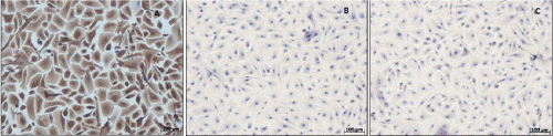
| Figure 1: Immunocytochemical analysis for quality control of cultured urothelial cells. In vitro staining of characteristic markers for (A) urothelial cells (AE1/AE3), (B) fibroblasts (TE-7), and (C) smooth muscle cells (1A4) demonstrated a pure HUC population without cellular contaminants indicated by lacking brownish colouration following antigen binding. |