|
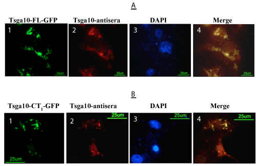
| Figure 4:Immunolocalization of the TSGA10 protein by transfection the COS-7 cells using full-length TSGA10-GFP (A) and TSGA10-CT1-GFP (B) constructs. By immunofluorescence, TSGA10 antibody detects specifically the TSGA10 protein (red, panels 2 in A&B) fused to GFP (green, panels 1 in A&B) and then merged (panels 4 in A&B). DAPI (blue, panels 3 in A&B) used for the nuclei staining. In each of A and B figures, several examples of overexpressed full-length and Carboxyl terminus (CT1) TSGA10 are presented. |