|
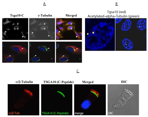
| Figure 5:Association of TSGA10 with the centrosome and basal body. A) Double immunofluorescence stain using antisera against TSGA10-C (left panel), anti-γ-tubulin (middle panel), and merged images (right panel) shows co-localization of these two proteins at a distinct cellular spot that seems to be associated with centrosomes. The experiments were carried out in COS-7 (top, x400; bottom, x1000) cells cultured on coverslips. DAPI (blue) was used to visualise nuclei. Immunostained COS- 7 cells are filtered for a better presentation (top panel), while TSGA10 and γ-tubulin are stained in red and green (bottom panel), respectively. B) Merged images of double immunofluorescence stain using antisera against TSGA10 and anti-γ-tubulin show co-localization (yellow) of these two proteins at two cellular spots that seems to be centrosomes. C) Sublocalization of TSGA10 in respiratory epithelial cells. Co-staining with antibodies against rabbit anti-a/b tubulin (red) and mouseanti- TSGA10 antibodies (green). a/b tubulin localizes to the entire length of the ciliary axonemes. In respiratory cells TSGA10 localizes to the ciliary base, which is consistent with localization at the basal bodies and the transition zone. |