 |
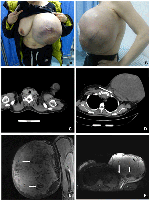
| Figure 1: (A) Anterorview of the breast tumor on presentation to our clinic; (B) Lateralview of the breast tumor on presentation to our clinic; (C,D) CT showed the chest wall was not involved (indicated by arrow (E,F) MR showed the giant tumor accompanied by multiple cystic part (indicated by arrows 1). Boundaries are still clear between the giant tumor and pectoralis (indicated by arrow 2). |