 |
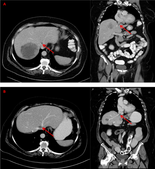
| Figure 1: CT images axial and coronar: (A) Before surgical therapy for the rectal carcinoma and liver metastases. (B) Two years after right-sided hemihepatectomy. These images show metastases in remaining segment one and four as well as a small mass between left and middle liver vein, which was suspicious for a liver metastasis. After surgical exploration and resection histopathological examination gave evidence that the latter was a subcapsular BC inside the liver parenchyma (red arrows). |