|
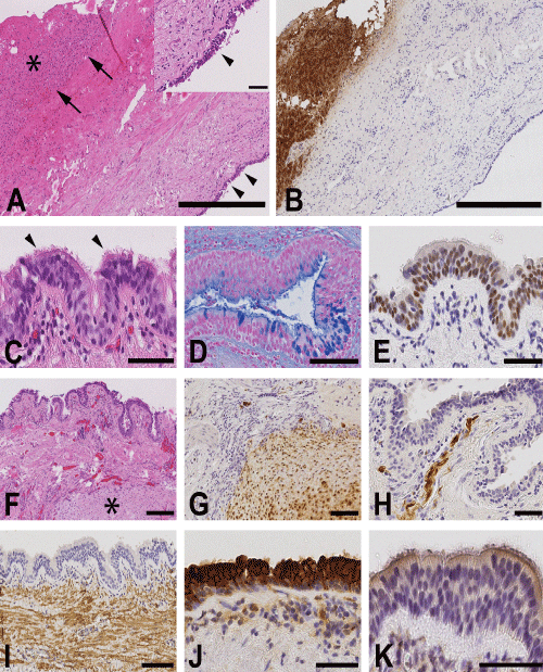
| Figure 2: Histological characterisation of the cystic lesion, showing inner surface of the cystic structures with morphological resemblance of bronchial mucosa and stroma with partial chondroid differentiation. A) Overview of the cyst with lining capsule and adjacent normal liver tissue. Arrows mark the border between capsule of the cyst and normal liver tissue. Arrow heads show lining epithelium of the cyst. Asterisk marking normal liver tissue. Bar: 500 μm. Inset: Magnification of the cyst lining epithelium. Bar: 50 μm. B) Immunostaining of hepatocytes. Bar: 500 μm. C) Cyst lining epithelium with cilia (arrow head). Bar: 50 μm. D) Alcian blue staining shows goblet cells. Bar: 100 μm. E) The cyst lining epithelium is positive for TTF-1. Bar: 50 μm. F) Overview of the adjacent stromal structures. Asterisk marking partial chondroid differentiation. Bar: 200 μm G) The adjacent chondroid differentiation is positive for S-100. Bar: 100 μm. H) S-100 immunostaining marks positive nerve fibres in the adjacent submucosal stroma. Bar: 50 μm. I) Immunostaining for smooth muscle actin. Bar: 100 μm. J) The cyst lining epithelium is positive for cytokeratin 7. Bar: 50 μm. K) The cyst lining epithelium is negative for cdx2. Bar: 50 μm. |