 |
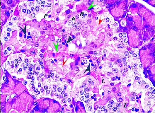
| Figure 2: A section from the diabetic group 1 (G2.1) pancreas showing an islet with loss of the normal architecture. The cells show degenerative changes in the form of vacuolated cytoplasm and swollen nuclei (stars) and cells with necrotic changes in the form of vacuolated cytoplasm with karyolytic nuclei (L), or karyorrhectic nuclei (R), dark eosinophilic cytoplasm with pyknotic nuclei (P) and complete loss of cells (red arrow). It also shows dilated congested capillaries (green arrow) and lymphocytic infiltration (black arrow) (H&E x1000). |