|
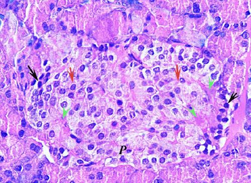
| Figure 4: A section from MSCs treated group (G3.1) pancreas showing cord arrangement of β-cells (red arrow) and Alpha cells at the margins of the islet (black arrow). Few cells with pyknotic nuclei and dark eosinophilic cytoplasm are seen (P). It also shows dilated congested blood capillaries (green heads) (H&E x1000). |