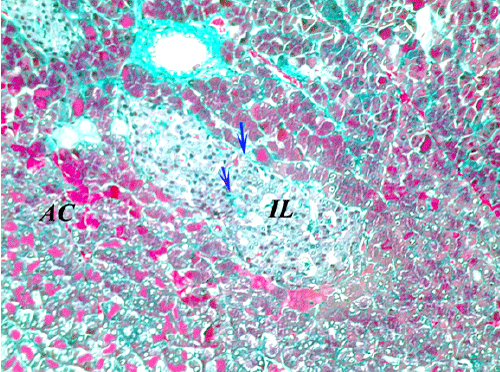
Figure 8:
A section from
control group
(G1) pancreas showing the normal distribution of collagen fibers, surrounding the islet and extending between its cells (blue arrow) (Masson’s Trichrome x400).