 |
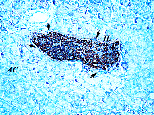
| Figure 13: A section from control group (G1) showing an islet (black arrows) surrounded by pancreatic acini. The cells in the core of the islet show a strong brown positive staining of their cytoplasm, with negative reaction in their nuclei. This reaction indicates the presence of the insulin secreting β-cells which are the major cells in the islet and are surrounded by a thin rim of Alpha cells which show a negative reaction. (Insulin antibody-6 Immunostain x400). |