|
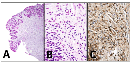
| Figure 1: Polypectomy specimen shows an onionskin-like concentric formation of spindle cells and dense inflammatory cell infiltration, predominantly eosinophils (hematoxylin and eosin, A, x20, B, x400). On immunohistochemistry (C, x400), the spindle cells appear diffusely positive for PDGFRA. |