|
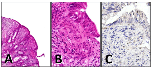
| Figure 2: Resected specimen shows striform- or whorl-like formation of spindle cells with abundant collagenous elements and eosinophilic cells (hematoxylin and eosin, A, x20, B, x400). On immunohistochemistry (C, x400), the spindle cells are negative for PDGFRA. |