 |
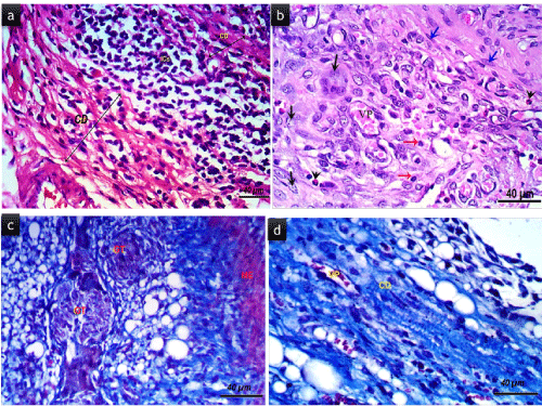
| Figure 3: Histological sections (stained with H&E X400 bar = 40 μm). (a) showing severe inflammatory cell infiltrate from group C. (b) invasion of different inflammatory cells primarily polymorph nuclear cells, macrophages( black arrows), plasma cells (red arrows), monocytes (head arrows) and red blood cells filled capillaries (VP) from group E. (c) increase in the number of the foreign body granulation tissue composed of spindle-shaped fibroblasts, new capillaries, dense inflammatory infiltrate and multinuclear giant cells (GT) in group C. (d) foreign body granulation tissue is absent in section of and dilated (VP) micro vessels group E. |