 |
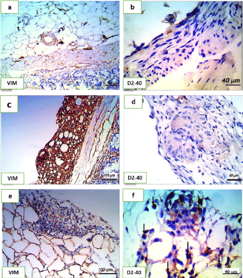
| Figure 5: Shows comparison between control, saline treated and garlic oil treated groups according to vimentin and D2-40 immunostaining reactions. (a) Mild brown staining in all layers (positive control) no signs of fibroblastic proliferation in group A. (b) Normal staining of the mesothelial layer, (white arrows), D2-40 immunostained section of group A, (c) Strong brown staining of the fibroblastic proliferative tissue in group C, (d) Negative cytoplasmic staining reaction in adhesive tissue, in group C. (e) Mild immunostaining expression of vimentin in mild fibro vascular tissue from group D. (f) Strong cytoplasmic immunostaining detecting mesothelial cells proliferation (arrows). |