profiles were presented here using Peak ScannerTM Software v1.0.
|
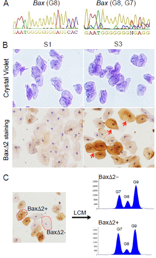
| Figure 1: Detection of Bax microsatellite mutation and BaxΔ2 isoform protein expression in human buccal cells. (A) Genomic DNA sequence of buccal cell samples from Control-1 (left) and Patient-1 (right) individuals. (B) Top, Crystal Violet staining of buccal cells from Control-1 and Patient-1 individuals; bottom, duplicate slides were immunohistochemically (IHC) stained with anti-BaxΔ2 antibody (2D4) [6]. The arrows indicate positive BaxΔ2 stained cells. (C) BaxΔ2- positive (BaxΔ2+) and -negative (BaxΔ2-) cells were isolated from Patient-1 IHC stained slide using a Zeiss PALM Laser Capture Microdissection System (LCM). The LCM captured cells (n=15 for each group) were subjected to genomic DNA isolation and fluorescence PCR with Bax primers covering Bax exon 3. The peak profiles were presented here using Peak ScannerTM Software v1.0. |