|
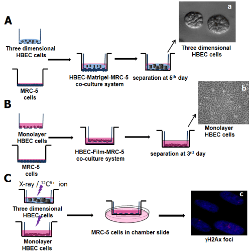
| Figure 1: A. The 3D HBEC-3KT organotypic culture model. (a): Representative confocal microscopic image of a 3D cyst culture when HBECs were cultured for 5 days in extracellular matrix and on the top of the feeder cells. B. The monolayer HBEC-3KT cell culture model. (b): Representative microscopic image of monolayer culture HBECs. C. The processes of monolayer and 3D culture irradiation and co-culture treatment. (c): Representative images of γH2AXfoci (red) formation in the blue nucleus region (DAPI staining) of the recipient MRC-5 cells co-cultured with the sham control and irradiated monolayer or 3D cultures |