|
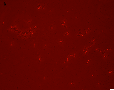
| Figure 3: Fluorescence microscopic integrity of artificial cells seeds that were prepared with Sph-DNA from E. coli -adenosine. The seeds that were prepared in Figure 2 are shown under a fluorescence microscope. Fluorescence was observed on the seeds. The image is the same field of view as shown in Figure 2, this image shows that the seeds possess DNA. Scale bar is 20 μm. |