|
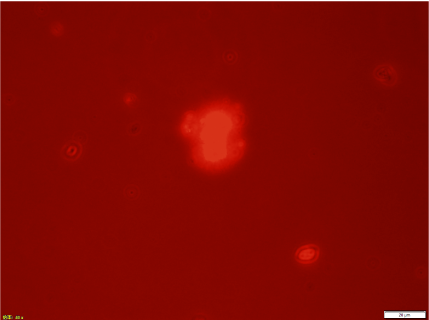
| Figure 6: Fluorescent microscopic image of seeds that were prepared with Sph-DNA from human placenta and F-fraction. Russet light is observed in each cell or a part of a cell aggregate, indicating that DNA is present in the seeds. The image is the same field of view shown in Figure 5. Scale bar is 20 μm. |