|
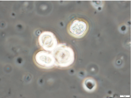
| Figure 7: Phase contrast microscopic image of artificial cells that were prepared with seeds (Sph-DNA from human placenta and F-fraction). Seeds were incubated in egg white for 7 days; after which, 1 ml of egg white was cultivated in D-MEM for 2 days. The aggregates were viewed under a phase contrast microscope. Each artificial cell had a roughly round or ellipse-like shape. Individual artificial cells and clusters of three cells were observed. Scale bar is 20 μm. |