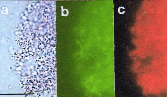
Figure 8: Fluorescent microscopy of artificial cells that were prepared with
seeds (Sph-DNA (E. coli plasmid) and H-extract) Sph solution was added to
E. coli DNA (plasmid). Then H-extract was added to the mixture. The mixture
(containing seeds) was incubated in egg white for 7 days. Egg white that
contained artificial cells was cultivated in D-MEM medium, and the aggregates
that formed at the bottom of each tube were stained with ethidium bromide
solution. The cells were smeared on a slide glass to form a cell monolayer.
Images a though c all show the same field of view.
a) Phase contrast microscope image without filters. Scale bar 20 μm.
b) Green light is observed in cell monolayer with the B: U-MNIBA2 filter,
indicating that GFP (green fluorescent protein) was present. Scale bar is
20 μm.
c) Russet light is observed in the cell monolayer with the G-U-MWIG2 filter,
indicating that DNA is present. Scale bar is 20 μm. |
