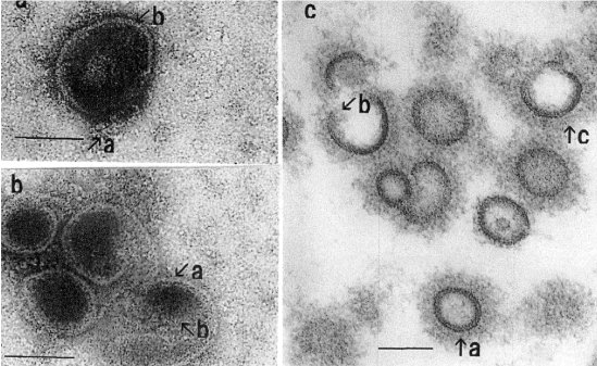
a) Negative scanning electron micrograph of an artificial cell. The cell is spherical (arrow a), and appears to be in an envelope (arrow b) Scale bar is 100 nm.
b) Negative scanning electron micrograph of artificial cell cluster. Artificial cells formed a cluster comprising four cells. One cell (arrow a) has an irregular shape with narrowing in the middle (arrow b). Scale bar is 100 nm.
c) Scanning electron micrograph of thin section of artificial cells. Artificial cells appear as rings (arrow a). One cell (arrow b) with a cut ring shape is also observed. These cells are coated with structures of irregular lengths (arrow c). Scale bar is 100 nm.