|
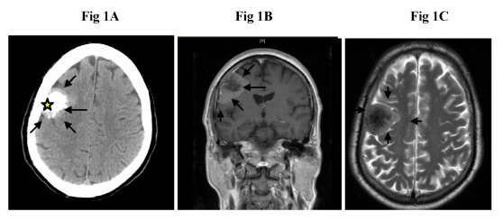
| Figure 1: A) The CT scan reveals the presence of a 3.1 cm subcortical tumor(indicated by arrow) located in the posterior frontal lobe of the right side. There is heavy calcification within the tumor (indicated by star). B) The extraaxial well-defined lobulated tumor with s relative mixed hypo- and isosignal intensity occupies the right temporal region. Increased signal density is in the periphery of the tumor (T1-weighted MRI). C) Decreased signal density in the central portion of tumor in the T2-weighted MRI. |