|
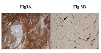
| Figure 3: A) Diffuse and strong cytoplasmic staining for EMA (brown color) (Original magnification: immunohistochemical staining by ABC method, 100x). B) CD34 staining highlights the rich vasculature in the background of tumor (Original magnification: immunohistochemical staining by ABC method, 400x). |