 |
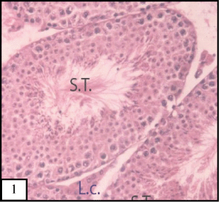
| Figure 1: Micrograph of testis section of mice in control group showing normal architecture of somniferous tubules (S.T.) A typical appearance of mice seminiferous tubules with different cellular association .The tubules had normal progression cells and Leydig cells have normal disruption (L.C.) (H&E., 200X). |