 |
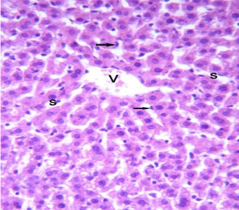
| Figure 5: A photomicrograph of the liver of control rats stained with H&E showing normal hepatic lobule has a thin walled central vein (V), hepatic cords radiating towards the periphery alternating with hepatic sinusoids(S) lined by Kupfer cells and endothelial cells (arrow). X, 400. |