|
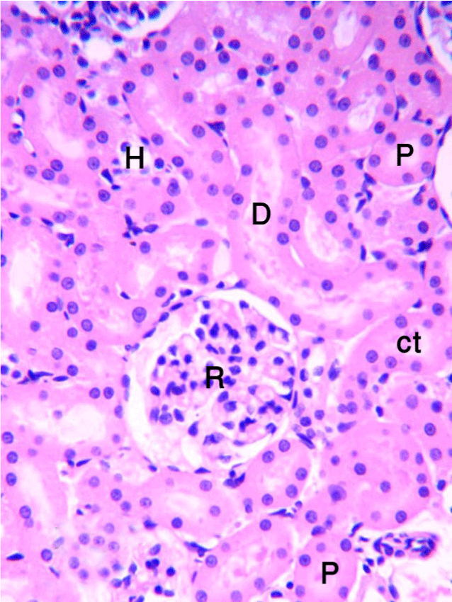
| Figure 17: A photomicrograph of the kidney of control rats stained with H&E showing normal histological structure of renal; corpuscle (R), proximal convoluted tubule (P), distal convoluted tubule (D), henles loop (H), and collecting tubule (CT). X, 400. |