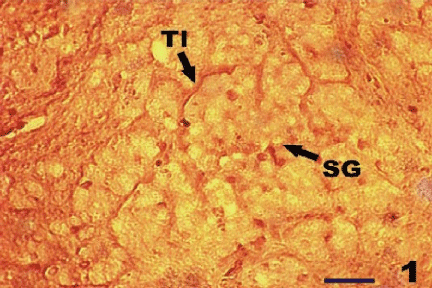
Figure 1:
Germinal epithelium of the testicular lobules (Tl) (arrow) and Spermatogonia (SG) showing diffuse and granular β-glucuronidase staining in resting phase of
L. rohita
X200.