|
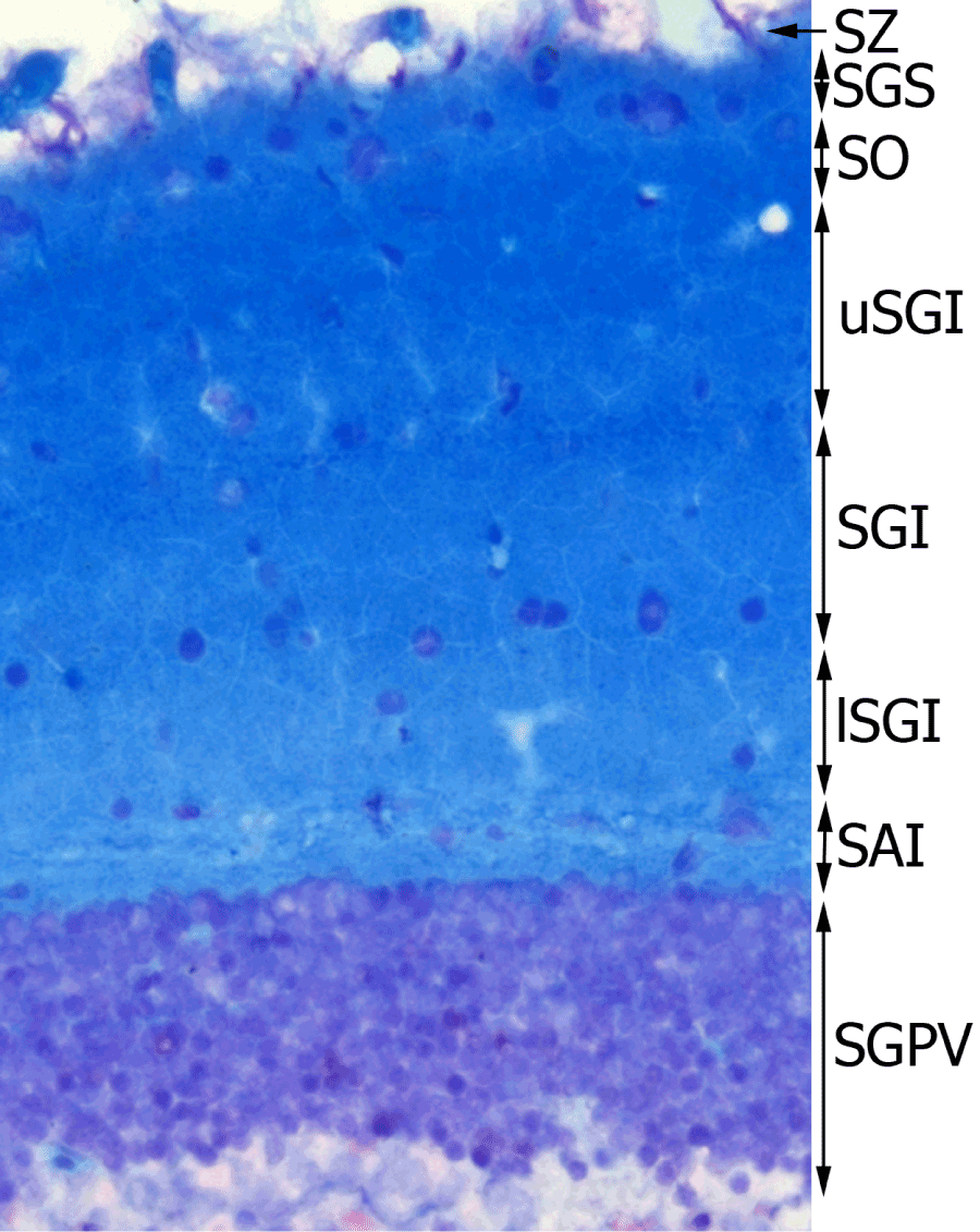
| Figure 1: 5µm fixed section of optic tectum of Nothobranchius guentheri stained with Luxol Fast Blue and Cresyl Violet. Optic tectum layers are labeled on the Figure: SZ, stratum zonale; SGS, stratum griseum superficiale; SO, stratum opticum; uSGI, upper stratum griseum intermediale; SGI, stratum griseum intermediale; lSGI, lower stratum griseum intermediale; SAI, stratum album intermediale; and SGPV, stratum griseum periventriculare. Neuronal cell bodies appear purple and myelin light blue. 400× magnification. |