 |
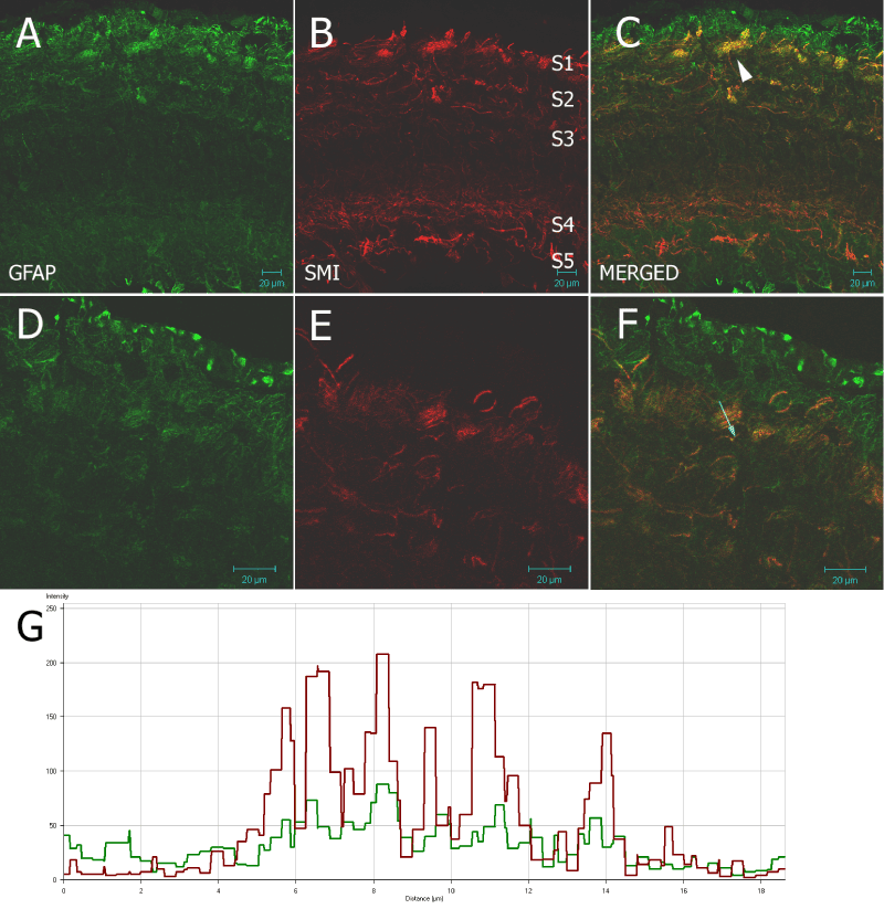
| Figure 3: 20 µm frozen section of optic tectum of Nothobranchius guentheri probed with rabbit anti-cow GFAP and with SMI31 antibodies. There is a strong GFAP positive layer corresponding with the SZ into the SO (Figure 1) which is indicative of radial glia. The SMI31 signal is seen in five discrete zones: S1 the SGS & SO; S2 the uSGI; S3 the SGI; S4 the SAI; and S5 the SGPV. SMI31 and anti-GFAP signal are seen to overlap in cage-like structures in the SGS which are enlarged in Figures D–F. There is some co-localization of SMI31 and anti-GFAP signal (Figures F & G) in these structures. Scale bar = 20µm. |