|
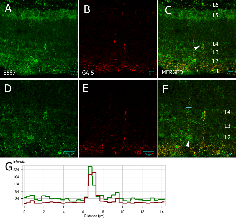
| Figure 6: 20 µm frozen section of optic tectum of Nothobranchius guentheri probed with E587 antiserum and mouse anti-human GFAP, GA-5. Arrow in (C) indicates a radial glial fiber which is the focus of Figures (DF). Six anti-L1 layers can be discerned. Arrow in (F) indicates a large E587 cell body, most likely a neuron. L1 corresponds with the SZ; L2 with the SGS/SO; L3 and L4 with the fiber tracts in the uSGI and SGI; L5 with the fiber layer of the SAI; and L6 with the SGPV. (G) Radial fiber in (F) bisected showing that green and red signal colocalize in the radial glial fiber spanning the OT. Scale bar represents 20 µm. |