 |
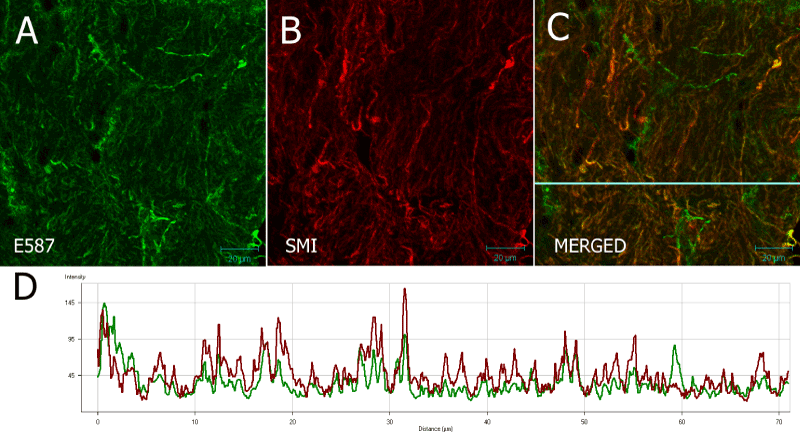
| Figure 7: 20 µm frozen section of optic nerve of Nothobranchius guentheri probed with rabbit E587 antiserum and SMI31. (C) shows a merged image showing co-localization (D) of red and green signal in some of the nerve fibers. Some fibers show no red signal and are probably astrocytic processes. Scale bar = 20 µm. |