|
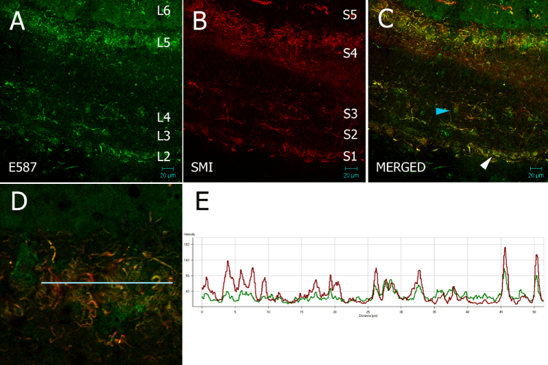
| Figure 8: 20 µm frozen section of optic tectum of Nothobranchius guentheri probed with rabbit E587 antiserum and mouse SMI31. Several discrete SMI31 and E587 layers are evident in (A) and (B) which partially overlap in the merged image (C). Blue arrow in (C) indicates a large E587 cell body, possibly a neuron. White arrow in (C) serves as the centre of focus for (D). Red and green signal in (D) colocalize (frame E) and show a complex of SMI31 and E587 positive fibers in the SGS/SO layer. |