 |
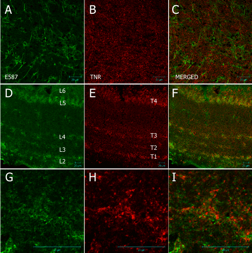
| Figure 9: 20 µm frozen section of optic nerve and optic tectum of Nothobranchius guentheri probed with rabbit E587 antibody mouse anti- TNR. (A–C) shows images of the ON. E587 positive astrocytic processes are visible along with anti-TNR signal which appears restricted to specific cell bodies. There is no evidence for colocalization as in Figures 5 and 7. (D–F) shows images of the OT. The anti-TNR signal (E) is ordered in four layers corresponding with the SO (T1); uSGI (T2); SGI (T3); and SAI (T4) into the SGPV. These layers appear broader than the corresponding E587 layers and each layer fades in the direction of the inner layers of the tectum. (G–I) show magnified images of the OT which clearly show that E587 and anti-TNR signal do not colocalize. The anti-TNR signal is restricted to small cell bodies in the tissue. Scale bar = 20 µm. |