 |
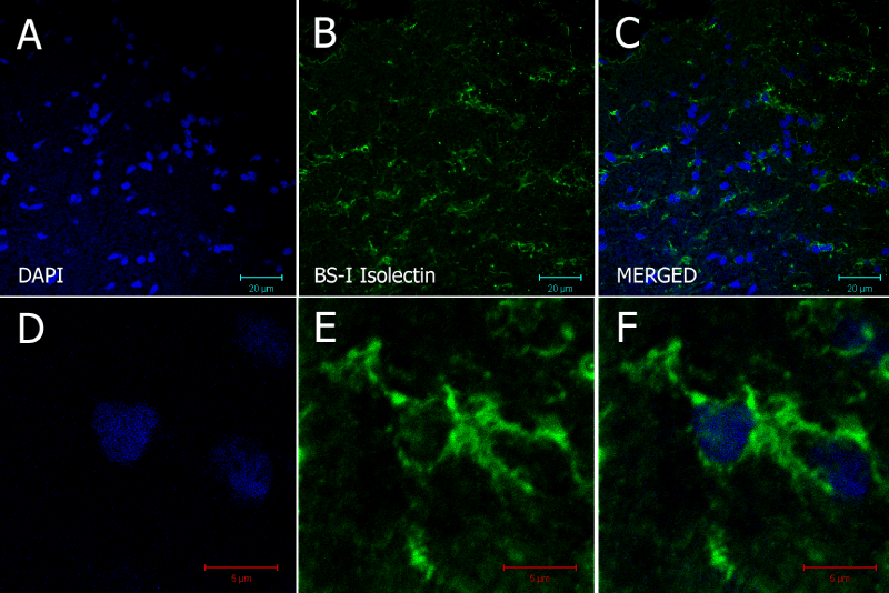
| Figure 10: 20 µm frozen section of optic nerve of Nothobranchius guentheri probed with DAPI and biotinylated BS-I Isolectin B4. (A–C) shows images of the ON containing microglia labeled with GS-isolectin. Typical microglial morphology can be observed in the enlarged frames (D–F). Scale bars = 20 µm for (A–C) and 5 µm for (D–F). |