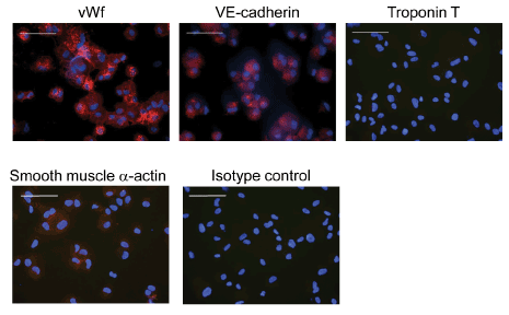
Figure 3
: Fluorescence microscopy illustrates that the clonally expanded hEHPCs were positive for endothelial cell markers vWf and VE-cadherin (red) but not for cardymyocyte marker troponin T or smooth muscle -actin. Bars indicate 100 μm.