|
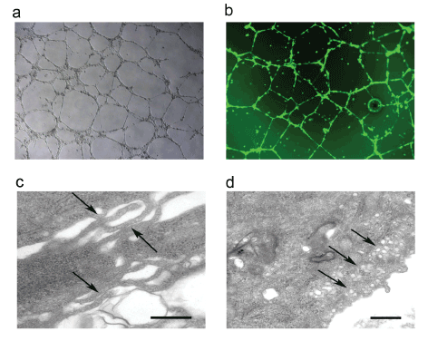
| Figure 4: a) Phase-contrast microscopy analysis of hEHPCs show differentiation into tubular structures on matrigel before transduction with GFP lentivirus and b) Fluorescence microscope analysis of hEHPCs differentiated into tubular structures on matrigel after transduction with GFP lentivirus. c) Electron microscopic analysis of isolated hEHPCs, representative pictures of transmission electron microscopy (TEM) of hEHPCs capillary-like structures in matrigel showing attachment between microvillus structures of endothelial like cells resembling tight junctions formation (arrows, scale bar; 0.5μm), and d) TEM image showing an hEHPCs plasma membrane with abundant pinocytotic vesicles (arrows, scale bar; 0.5μm). |