|
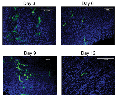
| Figure 5: Confocal microscopic analysis of SCID mouse heart. Representative images showing in vivo engraftment of GFP lentivirus transduced hEHPCs (green cells) in SCID mouse heart. Nuclei of cells are stained with DAPI, blue in colour. Cells could be traced until day 12 after transplantation. Shown are representative data from two independent experiments after transplanting the two hEHPCs isolated clones. Sections are 100 μm in thickness. |