|
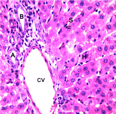
| Figure 1: Light photomicrography of liver of a control rat: the organ It is composed of lobules which are roughly hexagonal in shape, with portal triads at the vertices and a central vein (CV) in the middle. Within each lobule, hepatncytes (H) are arranged into hepatic cords running radiantly from [he central vein and are separated by adjacent blood sinusoids (S) containing Kupffer cells. N.B; Bile duct (B), Hepatic artery (A). H&E. x400 |