|
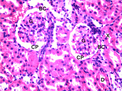
| Figure 3: Light photomicrography of Kidney (cortical part) of a control rat. The renal glomeruli (G) show normal structure and the proximal (X) are lined with typical thick cubic epithelium and distal (D) convoluted tubules are lined with the relatively low simple cubic epithelium. Organization of the glomeruli and a flat epithelium lining the glomerular capsule (BC) with distinct capsular space (CP) can be seen. H&E, x400. |