|
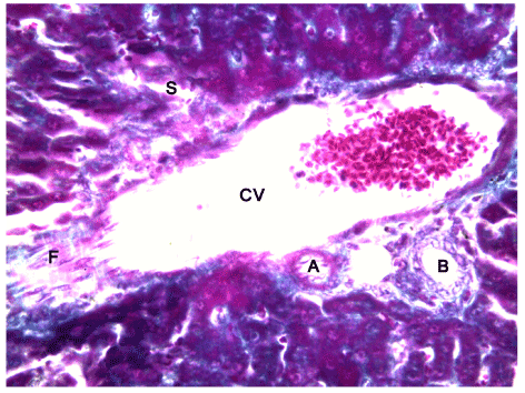
| Figure 6: Light photomicrography of liver of a rat after eight weeks of exposure to Cd Cl2. Showing (i): tight cytoplasm, enlarged cell sizes. Condensed nuclear chromatin. Sinusoidal widening (S) with accumulation of mononuclear cells in its vicinity, (ii): portal fibrosis (F).N.B; Bile duct (B). Central vein (V) and hepatic artery (A). Mallory’s x400. |