|
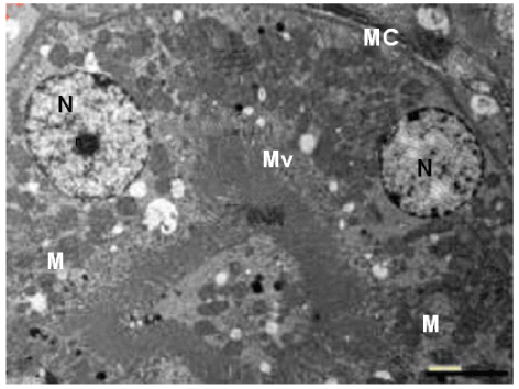
| Figure 20: Transmission electron microscopic picture of a section of kidney of control rat showing, the cells of the proximal convoluted tubule with euchromatic nuclei(N) and prominent nucleolus (n),many mitochondria (M) having normal crestae. The brush border of the cells has normal microvilli (Mv),and the basal lamina (CM) appears normal. TEM mag. =6000X. |