|
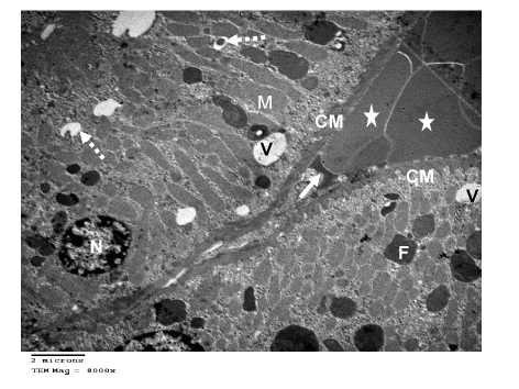
| Figure 21: Ultramicrograph of a section of liver of rat exposed to Cd Cl2 for 8 weeks , showing two liver cells with heterochromatic nuclei (N), numerous elongated mitochondria (M).Some lipid droplets (F),many lysosomes (broken arrows)as well as many vacuoles (V) appeared in cells cytoplasm,. A blood sinusoid (arrow) contains RPCs (*) is seen in between. The basal lamina appeared thick and infolded (CM). TEM mag. =8000X. |