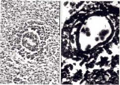
Figure 3:
Tissue sections showing (a) Angiocentric distribution of Lymphoma cells (H & E stain 100x), (b) Angionvasive lymphoma cells, locked within concentric rings of reticulin fiberes (Reticulin stain, 400x).