 |
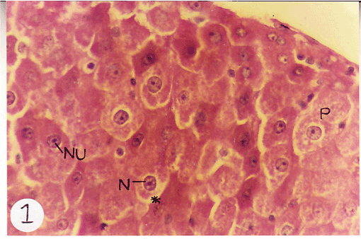
| Figure 1: Light Micrographs of rat liver treated with Cisplatin for 8 weeks shows the prescence of hepatocytes with increased vacuolation (*) having irregular morephology. Also note the damaged parenchyma (P) and large vacuolations around the nucleus (N). Almost all the nuclei posses prominent nucleolus (Nu). The photos are taken at the magnification (x 20). |