|
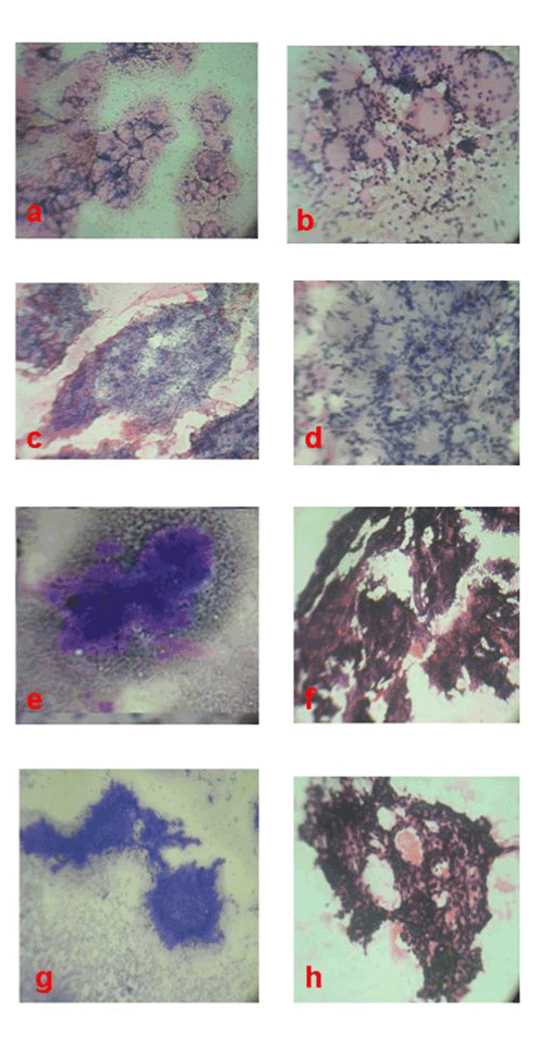
| Figure 1: Cylindroma. (a and b) Smears showing islands of epithelial cells in a palisaded manner (H and E,x100). and thick hyaline bands (H and E, x 400). Chondroid syringoma (c, d and e) Smears showing bland basaloid cells scattered in a fibrillary chondroid stoma (H and E,x100, x 400, MGG,x100). Papillary eccrine Adenoma.(f, g and h) Smears showing papillary fragments of bland basaloid cells fibro vascular core (H and E,x100, MGG,x100, H and E,x400). |