 |
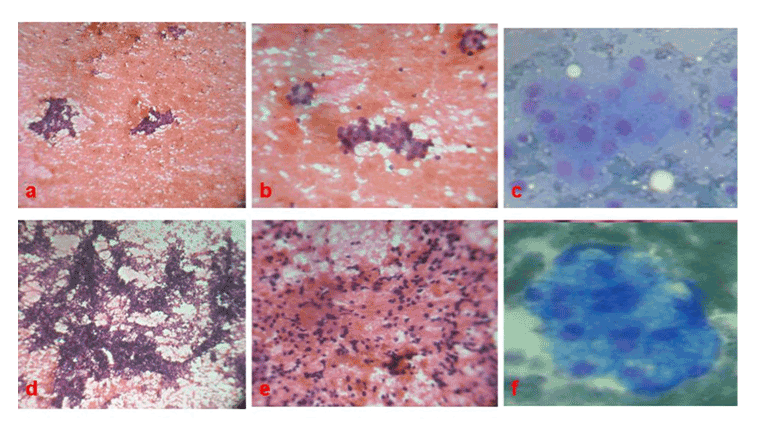
| Figure 2: Eccrine Spiradenoma. (a, b and c) Smear showing uniform cuboid cells in chusters and rosette formation (H and E,x100, H andE, x 400, MGG x 400) Nodular Hidradenoma (d, e and f) Smear showing tight clusters of a mixture of small round cells with clear cytoplasm and polygonal cells with basophilic cytoplasm (H and E x 100, H and E x 400, MGG x 400 |