|
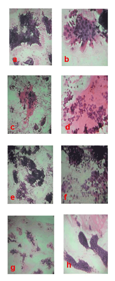
| Figure 3: Sweat gland carcinoma. (a and b) Smear showing pleomorphic epithelial cells in clusters and glandular structures (H and E x 100, x 400) Mucinous carcinoma of sweat gland. (c and d) Smear showing clusters of epithelial cells having mild nuclear pleomorphism in a lake of mucin (H and E x 100, x 400) Digital papillary Adenocarcinoma.(e and f) Smear showing syncytial aggregates and folding sheets of pleomorphic epithelial cells traversed by fibro vascular core (H and E x 100, x 400) Cutaneous adenoid cystic carcinoma. (g and h) Smear showing tridimensional globoid clusters of pleomorphic basaloid cells and acellular basement membrane (H and E x 100, x 400) |