|
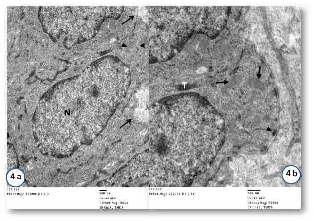
| Figure 4: Electron micrograph of Sham tongue basal cells showing (a) regular basal lamina (arrows), oval nuclei (N) with normal chromatin, regular nuclear membrane, abundant normal mitochondria (arrow heads) (Mic. Mag. X 3000). (b) Higher magnification of the previous showing basal cells, hemidesmosomes (arrow head), mitochondria (arrows) and abundant tonofillaments (T) (Mic. Mag. X 5000). |