|
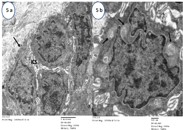
| Figure 5: Electron micrograph of OVX+P group epithelial cells showing (a) Irregular basal lamina (arrow), deformed nuclei with condensed and peripheralized chromatin and wide intercellular spaces (ICS ) (Mic. Mag. X2500). (b) Prickle cells showing distended and degenerated mitochondria (arrows) and decreased aggregation of tonofilaments (Mic. Mag. X5000). |